Back to Profile Page

Tree of Life Media Contributed By Jiri Vavra

<< < 1 2 > >>
ID Thumbnail Media Data
30014
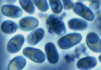
Scientific Name Glugea anomala
Comments fresh spores isolated from the tissue cyst of the stickleback. Many fish microsporidia have a very large vacuole in their spores.
Specimen Condition Live Specimen
Life Cycle Stage spores
Copyright © 2004 Ronny Larsson
Image Use ToL use only
Attached to Group Glugea (Microsporidia): view page image collection
Title glugea1.jpg
Image Type Photograph
Image Content Specimen(s)
ID 30014
30158
Scientific Name Binucleata daphniae
Comments spores(about 5.0 x 2.5 μm in size) isolated from infected hypodermal cells of infected Daphnia. Each spore shows a vacuole-like area in the posterior pole of the spore. This is a specific character of microsporidian spores.
Specimen Condition Live Specimen
Life Cycle Stage spores
Copyright © 2008
Image Use restricted
Attached to Group Binucleata (Microsporidia): view page image collection
Title binucleatasporynom2.jpg
Image Type Photograph
Image Content Specimen(s)
Technical Information Nomarski phase contrast micrograph
ID 30158
31008
Scientific Name Caudospora sp.
Comments fresh spore isolated from the fat body of Prosimulium mixtum (Diptera: Simuliidae). The spores of aquatic microsporidia bear often surface ornamentations, probably serving as floating devices.
Specimen Condition Live Specimen
Life Cycle Stage spore
Copyright © 1979
Image Use ToL use only
Attached to Group Caudospora (Microsporidia): view page image collection
Title caudospora.jpg
Image Type Photograph
Image Content Specimen(s)
ID 31008
30995
Scientific Name Chytridiopsis typographi
Specimen Condition Dead Specimen
Life Cycle Stage spore
Copyright © 2008
Image Use restricted
Attached to Group Chytridiopsis (Microsporidia): view page image collection
Title chytrdsporaexcelentni.jpg
Image Type Photograph
Image Content Specimen(s), Ultrastructure
Technical Information TEM micrograph
ID 30995
30996
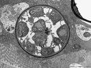
Scientific Name Chytridiopsis typographi
Specimen Condition Dead Specimen
Life Cycle Stage thickwalled spore sac with young spores
Copyright © 2008
Image Use restricted
Attached to Group Chytridiopsis (Microsporidia): view page image collection
Title chytridcystalepsi.jpg
Image Type Photograph
Image Content Specimen(s), Ultrastructure
Technical Information TEM micrograph
ID 30996
30998
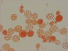
Scientific Name Vavraia culicis subsp. floridensis Vavra and Becnel 2007
Specimen Condition Live Specimen
Life Cycle Stage thin-walled sporophorous vesicles with spores
Copyright © 2007
Image Use restricted
Attached to Group Vavraia (Microsporidia): view page image collection
Title culicisspov40xfin.jpg
Image Type Photograph
Image Content Specimen(s)
Technical Information in vivo stained preparation (Congo Red)
ID 30998
30999
Scientific Name Marssoniella elegans Lemmermann 1900
Comments Spores of Marssoniella elegans occur in groups of four or eight and are surounded by thick mucous capsule for flotation.
Specimen Condition Live Specimen
Life Cycle Stage spore groups isolated from infected copepode host (Cyclops vicinus)
Copyright © 2005
Image Use restricted
Attached to Group Marssoniella (Microsporidia): view page image collection
Title marsson-spory.jpg
Image Type Photograph
Image Content Specimen(s)
Technical Information spores suspended in dilluted India Ink
ID 30999
31002
Scientific Name Brachiola algerae
Specimen Condition Dead Specimen
Life Cycle Stage developmental stage - meront (vegetative cell)
Body Part note two nuclei in diplokaryon configuration (N,N), atypical Golgi zone (G) and cytoplasm rich in ribosomes. The plasma membrane is covered with tubular glykocalyx material charateristic for the genus Brachiola.
Copyright © 2001
Image Use restricted
Attached to Group Brachiola (Microsporidia): view page image collection
Title meront-brachcopy.jpg
Image Type Photograph
Image Content Specimen(s), Ultrastructure
Technical Information TEM micrograph
ID 31002
31460
Scientific Name Brachiola algerae (formerly Nosema algerae)
Comments In fully extruded spore the posterior vacuole expanded filling the entire volume of the spore. The spore now is essentially empty and all its contents were pushed into the everting filament.
Copyright ©
Image Use restricted
Attached to Group Brachiola (Microsporidia): view page image collection
Title prazdnaspora1.jpg
Image Type Photograph
Image Content Ultrastructure
ID 31460
31745
Comments Composite image of various microporidian spore types.
Copyright ©
Image Use restricted
Attached to Group Microsporidia: view page image collection
Title velkespory.jpg
Technical Information Light microscope images of fresh spores. The centre image: scanning electron microscope image of spores of Glugea anomala.
ID 31745
Please note: Most images and other media displayed on the Tree of Life web site are protected by copyright, and the ToL cannot act as an agent for their distribution. If you would like to use any of these materials for your own projects, you need to ask the copyright owner(s) for permission. For additional information, please refer to the ToL Copyright Policies.

Contribute to the ToL

ToL Contributions

Ways to Contribute

Use of Contributions

Scientific Content

Building Treehouses

Images/Media

  go to the Tree of Life home page
top