Utegenia shpinari
Michel Laurin
Introduction
Utegenia shpinari was found in the Upper Pennsylvanian or Lower Permian of Kazakhstan (the stratigraphy of this area is problematic). It is represented by more than four hundred flattened but well preserved specimens. These specimens document a growth series ranging from small larvae to small post-metamorphic specimens. This growth series shows that the neural arches ossify before the centra, and the external gills disappear when the skull reaches a length of about 1.5 cm (Kuznetsoz and Ivakhnenko, 1981). However, the poor ossification of most endochondral elements suggests that no fully mature specimen was found. The cranial length of the known specimens ranges from 1 cm to 3.5 cm (Kuznetsov and Ivakhnenko, 1981).
Characteristics
The first published reconstructions of Utegenia shpinari showed a very broad skull (Kuznetsov and Ivakhnenko, 1981), but recent work suggests that Kuznetsov and Ivakhnenko did not take the extensive crushing into consideration (Fig. 1), and that the skull was somewhat more narrow (Laurin, in press). The braincase is virtually unknown because its endochondral elements were cartilaginous. However, the parasphenoid was broad, triangular, and covered in a shagreen of denticles.


Figure 1. Skull of a postmetamorphic specimen of Utegenia shpinari. Notice the contact between the postorbital (Po) and the supratemporal (St).
Utegenia had a moderately long trunk, with 28 presacral vertebrae (three of four more than in other seymouriamorphs). The neural arches are paired and disarticulated from the pleurocentra even in the largest known specimen, but this is probably a juvenile character (neural arch fusion occurred relatively late in the ontogeny of early terrestrial vertebrates).
The appendicular skeleton is poorly known because of its poor ossification. The scapula and coracoid are discrete elements, and the scapula is approximately circular. The ends of the limb bones are unossified. The carpus and tarsus are usually not ossified.
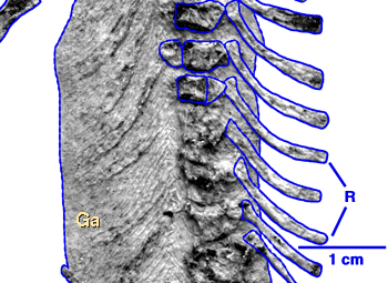
Utegenia had circular scales over most of its body, like Discosauriscus. It retained rhomboidal ventral scales (Fig. 2) arranged in a chevron pattern (gastralia) and a contact between the postorbital and the supratemporal (Fig. 1). Both of these characters suggest that it is not closely related to Discosauriscus, Ariekanerpeton, or Seymouria (none of these seymouriamorphs appear to have had rhomboidal gastralia, although circular ventral scales were present in Discosauriscus, and the presence of gastralia cannot be determined in Seymouria). The contact between the supratemporal and the postorbital is rarely present in Discosauriscus and Ariekanerpeton, and it is not found in Seymouria, but it is present in most postmetamorphic specimens of Utegenia.


Figure 2. Impressions of ventral scales of Utegenia.
References
Kuznetsov, V.V., and M.F. Ivakhnenko. 1981. Discosauriscids from the Upper Paleozoic in Southern Kazakhstan. Paleontological Journal 1981: 101-108.
Laurin, M. In press. A reappraisal of Utegenia, a Permo-Carboniferous seymouriamorph (Tetrapoda: Batrachosauria) from Kazakhstan. Journal of Vertebrate Paleontology 29 pages, 6 figures.
Title Illustrations

| Scientific Name | Utegenia shpinari |
|---|---|
| Location | Kazakhstan |
| Comments | Skeleton of a postmetamorphic specimen. This specimen is one of more than four hundred skeletons recently collected in Kazakhstan. Impressions of ventral scales are preserved on the right side of the abdomen. |
| Specimen Condition | Fossil |
| View | ventral |
| Image Use |
 This media file is licensed under the Creative Commons Attribution-NonCommercial License - Version 3.0. This media file is licensed under the Creative Commons Attribution-NonCommercial License - Version 3.0.
|
| Copyright |
© 1996 Michel Laurin

|
About This Page
I thank my wife (Ms. Patricia Lai) and Mr. Matthew Marlowe for editing this page. Dr. David Maddison provided invaluable assistance in formatting this page and in linking it to other pages of the Tree of Life. I thank Dr. Jozef Klembara for his useful comments on Discosauriscus.
Michel Laurin

Muséum National d'Histoire Naturelle, Paris, France
Correspondence regarding this page should be directed to Michel Laurin at
Page copyright © 1996 Michel Laurin
 Page: Tree of Life
Utegenia shpinari.
Authored by
Michel Laurin.
The TEXT of this page is licensed under the
Creative Commons Attribution License - Version 3.0. Note that images and other media
featured on this page are each governed by their own license, and they may or may not be available
for reuse. Click on an image or a media link to access the media data window, which provides the
relevant licensing information. For the general terms and conditions of ToL material reuse and
redistribution, please see the Tree of Life Copyright
Policies.
Citing this page:
Laurin, Michel. 1996. Utegenia shpinari. Version 01 January 1996. http://tolweb.org/Utegenia_shpinari/17542/1996.01.01 in The Tree of Life Web Project, http://tolweb.org/


 Go to quick links
Go to quick search
Go to navigation for this section of the ToL site
Go to detailed links for the ToL site